
We engineer cells and materials that communicate and process information through synthetic biology
Our inspiration is the ability of organisms and the materials they are made of to adapt to dynamic environmental conditions. Plants adapt growth to light conditions; bacteria develop resistance against antibiotics or bones get stronger when exercised. The basis for this ability to adapt is a fascinating information processing machinery of the organisms: Environmental conditions are captured by molecular sensors, then the signals are processed and integrated with genetic programs to finally yield a targeted response.
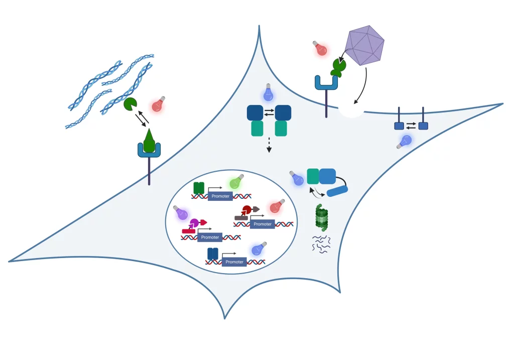
In our research, we engineer nature’s molecular sensing, processing, and actuation machinery in order to precisely control the function and properties of cells and materials. We apply these newly developed technologies in different fields of fundamental and applied research.

Team Members


Research
Stimulus-responsive and Information-processing (living) Materials

We develop and apply stimulus-responsive and information-processing biohybrid polymer materials. To this aim, we functionally couple synthetic biological molecular sensors and switches to polymer materials. By wiring these switches according to topologies inspired by electronic circuits, we engineer materials that perform fundamental computational operations. Examples of our work include:
- We engineered a hydrogel based on a bacteria-derived photoreceptor which allows the light-responsive, fully reversibly tuning of its mechanical properties. We applied this hydrogel as extracellular matrix to analyze the impact of dynamic mechanical environments on transcriptome-wide responses in mesenchymal stem cells or on the migration of T-lymphocytes.
See Hörner et al. Advanced Materials 2019 - We integrated synthetic biological switches with polymer materials into a circuit inspired by an electronic counter. The resulting material system was able to count the number of input light pulses and to release different output as a function of the number of light pulses detected. We applied this system to sequentially release different biocatalysts to drive a two-step biochemical reaction.
See Beyer et al., Advanced Materials 2018 - We developed PenTag, a protein tag for the spontaneous, covalent coupling of proteins to ampicillin-functionalized molecules such as dyes, polymers, or solid supports. Based on this strategy, we engineered and assembled material modules to function as encoder for processing different combinations of biochemical input stimuli.
See Mohsenin et al., Advanced Functional Materials 2024 - By engineering modular protease-based switches that can either be activated or repressed, we develop information-processing biohybrid circuits that process binary biomolecular information according to a circuit inspired by electronic decoders. Such circuits can be applied to process and interpret biochemical sensor information for advanced diagnostic applications.
See Mohsenin et al., Advanced Materials 2024
Molecular optogenetics to control cell fate and function
We develop and apply molecular optogenetic tools to control cell fate and function with unprecedented spatial and temporal precision in a dose-dependent and highly specific manner. To this aim, we engineer plant- and bacteria-derived photoreceptors and functionally couple them to proteins involved in cell signaling and gene expression. Examples of our work include:
- Light-inducible formation of liquid or gel-like transcription factor condensates in mammalian cells and mice. We demonstrate that liquid “transcription factor droplets” show a several-fold higher activity in inducing transgene expression compared to native transcription factors. Further, gel-like transcription factor condensates were shown to correlate with decreased transcriptional activation thus providing a materials-based layer of controlling gene expression.
See Schneider et al., Science Advances 2021 and Fischer et al., Small 2024 - Light-guided adeno-associated viral (AAV) vectors. We engineered a light-responsive tropism into AAVs which allows us to selectively transfer genetic information into single cells or to transduce different cells within one culture with different transgenes.
See Hörner et al., Science Advances 2021
Our group is running www.optobase.org, the most comprehensive database on molecular optogenetics. Have a look and discover the amazing opportunities in controlling biology with light!
Biosensors
We integrate natural and engineered molecular sensors for drugs, metabolites or nucleic acids into suitable readout formats for the fast and sensitive quantification of such substances. Together with collaboration partners, we develop biosensor systems for different application fields:
Open Positions
We are always excited to meet curious and creative scientists passionate about synthetic biology, optogenetics, and engineered living materials. If you would like to shape the future of biobased and living materials with us, we warmly welcome your spontaneous application for a PhD thesis or Postdoc position!
Projects and Partners
We perform collaborative research in materials-oriented synthetic biology within interdisciplinary research consortia
STEADY
Within the ERC Advanced Grant STEADY, we develop concepts for dynamically controlling the properties of engineered living materials by advanced synthetic genetic circuits.
LoopOfFun
We coordinate the European Innovation Council (EIC)-funded consortium LoopOfFun in which we aim at developing a platform for the rapid development of industry-scale, one-step, simple casting-based manufacturing processes for fungal mycelia composites. We jointly work towards this goal with our consortium partners:
- Prof. Roman Jerala, National Institute of Chemistry, Ljubljana, Slovenia
- Dr. Achim Weber, Fraunhofer IGB, Stuttgart, Germany
- Prof. Arnold Driessen, University of Groningen, The Netherlands
- Carlotta Borgato and Jan Boelen, Atelier LUMA, Arles, France
DELIVER
In the project DELIVER funded by the Carl-Zeiss-Foundation, we collaborate towards the data-driven engineering of sustainable living materials. We combine synthetic biology with materials sciences and data-driven approaches to design bio-based composite materials with custom-tailored structural properties for construction applications. Within deliver, we collaborate with the following partners:
- Prof. Thomas Speck, University of Freiburg, Germany
- Dr. Clemens Kreutz, University Hospital Freiburg, Germany
BILLARD
We coordinate the BILLARD project funded by the Federal Ministry of Education and Research (BMBF) within the funding line “Biologization of Technology”, we collaborate with PD Dr. Felicitas Bucher from the Clinic of Ophtamology at the University Hospital Freiburg on the development of novel intraocular drug delivery devices.
CIBSS – Centre for Integrative Biological Signalling Studies
We are member of the Cluster of Excellence CIBSS in which we perform research on novel optogenetic technologies to control signaling reactions in mammalian cells. We mainly collaborate with Prof. Dr. Jens Timmer on the model-based design of synthetic biological switches and networks and with Prof. Dr. Wolfgang Schamel on controlling immunological processes such as T cell activation via optogenetics.
Publications
Beyer, H. M. | Engesser, R. | Hörner, M. | Koschmieder, J. | Beyer, P. | Timmer, J. | Zurbriggen, M. D. | Weber, Wilfried
DOI:
Synthetic biology applies engineering concepts to build cellular systems that perceive and process information. This is achieved by assembling genetic modules according to engineering design principles. Recent advance in the field has contributed optogenetic switches for controlling diverse biological functions in response to light. Here, the concept is introduced to apply synthetic biology switches and design principles for the synthesis of multi-input-processing materials. This is exemplified by the synthesis of a materials system that counts light pulses. Guided by a quantitative mathematical model, functional synthetic biology-derived modules are combined into a polymer framework resulting in a biohybrid materials system that releases distinct output molecules specific to the number of input light pulses detected. Further demonstration of modular extension yields a light pulse-counting materials system to sequentially release different enzymes catalyzing a multistep biochemical reaction. The resulting smart materials systems can provide novel solutions as integrated sensors and actuators with broad perspectives in fundamental and applied research. © 2018 WILEY-VCH Verlag GmbH & Co. KGaA, Weinheim
Beyer, H. M. | Thomas, O. S. | Riegel, N. | Zurbriggen, M. D. | Weber, Wilfried | Hörner, M.
DOI:
Molecular traps can control activity and abundance of many biological factors. Here, we report the development of a generic opto-trap to reversibly bind and release biomolecules with high spatiotemporal control by illumination with non-invasive and cell-compatible red and far-red light. We use the Arapidopsis thaliana photoreceptor phytochrome B to regulate the release of diverse proteins from a variety of material scaffolds. Fusion of a short 100 amino acids “PIF-tag” derived from the phytochrome interacting factor 6, renders arbitrary molecules opto-trap-compatible. Reversible opto-trapping of target molecules enables novel possibilities for future developments in diagnostics, therapeutics, and basic research. Statement of Significance: The investigation of cellular signaling events or the development of complex therapeutics and integrative diagnostic devices requires the deliberate control of biomolecule abundance and activity. During recent years, the use of natural photoreceptors within cells leveraged the control of diverse cellular events, benefiting from the superior spatial and temporal control characteristics of light as compared to conventional chemical stimuli. Concurrently, biological switches entailing intrinsic compatibility toward biological environments increasingly found application in biohybrid materials. We employ the plant red/far-red photoreceptor phytochrome B, which reversibly interacts with its phytochrome interacting factors (PIFs), for developing a generic opto-trap. This platform allows the use of red and far-red light to spatiotemporally control binding and release of arbitrary PIF-fused biomolecules from various material scaffolds. © 2018 Acta Materialia Inc.
Chatelle, C. | Ochoa-Fernandez, R. | Engesser, R. | Schneider, N. | Beyer, H. M. | Jones, A. R. | Timmer, J. | Zurbriggen, M. D. | Weber, Wilfried
DOI:
The ever-increasing complexity of synthetic gene networks and applications of synthetic biology requires precise and orthogonal gene expression systems. Of particular interest are systems responsive to light as they enable the control of gene expression dynamics with unprecedented resolution in space and time. While broadly used in mammalian backgrounds, however, optogenetic approaches in plant cells are still limited due to interference of the activating light with endogenous photoreceptors. Here, we describe the development of the first synthetic light-responsive system for the targeted control of gene expression in mammalian and plant cells that responds to the green range of the light spectrum in which plant photoreceptors have minimal activity. We first engineered a system based on the light-sensitive bacterial transcription factor CarH and its cognate DNA operator sequence CarO from Thermus thermophilus to control gene expression in mammalian cells. The system was functional in various mammalian cell lines, showing high induction (up to 350-fold) along with low leakiness, as well as high reversibility. We quantitatively described the systems characteristics by the development and experimental validation of a mathematical model. Finally, we transferred the system into A. thaliana protoplasts and demonstrated gene repression in response to green light. We expect that this system will provide new opportunities in applications based on synthetic gene networks and will open up perspectives for optogenetic studies in mammalian and plant cells. Copyright © 2018 American Chemical Society.
Hörner, M. | Chatelle, C. | Mühlhäuser, W. W. D. | Stocker, D. R. | Coats, M. | Weber, Wilfried | Radziwill, G.
DOI:
Focal adhesion kinase (FAK) integrates signaling from integrins, growth factor receptors and mechanical stress to control cell adhesion, motility, survival and proliferation. Here, we developed a single-component, photo-activatable FAK, termed optoFAK, by using blue light-induced oligomerization of cryptochrome 2 (CRY2) to activate FAK-CRY2 fusion proteins. OptoFAK functions uncoupled from physiological stimuli and activates downstream signaling rapidly and reversibly upon blue light exposure. OptoFAK stimulates SRC creating a positive feedback loop on FAK activation, facilitating phosphorylation of paxillin and p130Cas in adherent cells. In detached cells or in mechanically stressed adherent cells, optoFAK is autophosphorylated upon exposure to blue light, however, downstream signaling is hampered indicating that the accessibility to these substrates is disturbed. OptoFAK may prove to be a useful tool to study the biological function of FAK in growth factor and integrin signaling, tension-mediated focal adhesion maturation or anoikis and could additionally serve as test system for kinase inhibitors. © 2017
Kolar, K. | Knobloch, C. | Stork, H. | Žnidarič, M. | Weber, Wilfried
DOI:
OptoBase is an online platform for molecular optogenetics. At its core is a hand-annotated and ontology-supported database that aims to cover all existing optogenetic switches and publications, which is further complemented with a collection of convenient optogenetics-related web tools. OptoBase is meant both for expert optogeneticists to easily keep track of the field, as well as for all researchers who find optogenetics inviting as a powerful tool to address their biological questions of interest. It is available at https://www.optobase.org. This work also presents OptoBase-based analysis of the trends in molecular optogenetics. © 2018 American Chemical Society.
Middleton, A. M. | Dal Bosco, C. | Chlap, P. | Bensch, R. | Harz, H. | Ren, F. | Bergmann, S. | Wend, S. | Weber, Wilfried | Hayashi, K. I. | Zurbriggen, M. D. | Uhl, R. | Ronneberger, O. | Palme, K. | Fleck, C. | Dovzhenko, A.
DOI:
In plants, the phytohormone auxin acts as a master regulator of developmental processes and environmental responses. The best characterized process in the auxin regulatory network occurs at the subcellular scale, wherein auxin mediates signal transduction into transcriptional programs by triggering the degradation of Aux/IAA transcriptional repressor proteins in the nucleus. However, whether and how auxin movement between the nucleus and the surrounding compartments is regulated remain elusive. Using a fluorescent auxin analog, we show that its diffusion into the nucleus is restricted. By combining mathematical modeling with time course assays on auxin-mediated nuclear signaling and quantitative phenotyping in single plant cell systems, we show that ER-to-nucleus auxin flux represents a major subcellular pathway to directly control nuclear auxin levels. Our findings propose that the homeostatically regulated auxin pool in the ER and ER-to-nucleus auxin fluxes underpin auxin-mediated downstream responses in plant cells. Middleton et al. study how the plant phytohormone auxin enters the nucleus by using quantitative phenotyping in single plant cell systems and bespoke mathematical models that relate controlled perturbations to experimentally measurable responses. Their findings show that auxin predominantly enters the nucleus via the endoplasmic reticulum. © 2018 The Authors
Schneider, N. | Gäbelein, C. | Wiener, J. | Georgiev, T. | Gobet, N. | Weber, Wilfried | Meier, M.
DOI:
We here present a method that combines genetic code expansion with CRISPR/Cas9 genome engineering to label endogenously expressed proteins with high spatiotemporal resolution. The method exploits the use of an orthogonal tRNA/tRNA synthetase pair in conjugation with noncanonical amino acids to create stop codon read through events. To demonstrate the functionality of the method, we pulse labeled endogenous β-actin and tumor protein p53 with a minimally invasive HA tag at their C-termini. Targeting the protein label with a proximity ligation assay plus real time imaging facilitates seamless quantification of the protein synthesis rate and spatial localization at the single cell level. The presented approach does not interfere with any physiological control of cellular expression, nor did we observe any perturbation of endogenous protein functions. © 2018 American Chemical Society.
Wagner, H. J. | Engesser, R. | Ermes, K. | Geraths, C. | Timmer, J. | Weber, Wilfried
DOI:
The translation of engineering designs to materials sciences by means of synthetic biological tools represents a novel concept for the development of information-processing materials systems. Here, we provide data on the mathematical model-guided implementation of a biomaterials-based positive feedback loop for the detection of proteolytic activities. Furthermore, we present data on an extended system design for the detection of the antibiotic novobiocin. This work is related to the research article “Synthetic biology-inspired design of signal-amplifying materials systems” (Wagner et al., 2018) [1]. © 2018 The Authors
Wagner, H. J. | Wehrle, S. | Weiss, E. | Cavallari, M. | Weber, Wilfried
DOI:
Nanobodies, the smallest possible antibody format, have become of considerable interest for biotechnological and immunotherapeutic applications. They show excellent robustness, are non-immunogenic in humans, and can easily be engineered and produced in prokaryotic hosts. Traditionally, nanobodies are selected from camelid immune libraries involving the maintenance and treatment of animals. Recent advances have involved the generation of nanobodies from naïve or synthetic libraries. However, such approaches demand large library sizes and sophisticated selection procedures. Here, we propose an alternative, two-step approach for the design and generation of nanobodies. In a first step, complementarity-determining regions (CDRs) are grafted from conventional antibody formats onto nanobody frameworks, generating weak antigen binders. In a second step, the weak binders serve as templates to design focused synthetic phage libraries for affinity maturation. We validated this approach by grafting toxin-and hapten-specific CDRs onto frameworks derived from variable domains of camelid heavy-chain-only antibodies (VHH). We then affinity matured the hapten binder via panning of a synthetic phage library. We suggest that this strategy can complement existing immune, naïve, and synthetic library based methods, requiring neither animal experiments, nor large libraries, nor sophisticated selection protocols. © 2018 by the authors. Licensee MDPI, Basel, Switzerland.
Bruch, R. | Chatelle, C. | Kling, A. | Rebmann, B. | Wirth, S. | Schumann, S. | Weber, Wilfried | Dincer, C. | Urban, G.
DOI:
An appropriate antibiotherapy is crucial for the safety and recovery of patients. Depending on the clinical conditions of patients, the required dose to effectively eradicate an infection may vary. An inadequate dosing not only reduces the efficacy of the antibiotic, but also promotes the emergence of antimicrobial resistances. Therefore, a personalized therapy is of great interest for improved patients' outcome and will reduce in long-term the prevalence of multidrug-resistances. In this context, on-site monitoring of the antibiotic blood concentration is fundamental to facilitate an individual adjustment of the antibiotherapy. Herein, we present a bioinspired approach for the bedside monitoring of free accessible ß-lactam antibiotics, including penicillins (piperacillin) and cephalosporins (cefuroxime and cefazolin) in untreated plasma samples. The introduced system combines a disposable microfluidic chip with a naturally occurring penicillin-binding protein, resulting in a high-performance platform, capable of gauging very low antibiotic concentrations (less than 6 ng ml-1) from only 1 μl of serum. The system's applicability to a personalized antibiotherapy was successfully demonstrated by monitoring the pharmacokinetics of patients, treated with ß-lactam antibiotics, undergoing surgery. © 2017 The Author(s).