
We engineer cells and materials that communicate and process information through synthetic biology
Our inspiration is the ability of organisms and the materials they are made of to adapt to dynamic environmental conditions. Plants adapt growth to light conditions; bacteria develop resistance against antibiotics or bones get stronger when exercised. The basis for this ability to adapt is a fascinating information processing machinery of the organisms: Environmental conditions are captured by molecular sensors, then the signals are processed and integrated with genetic programs to finally yield a targeted response.
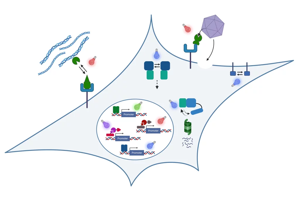
In our research, we engineer nature’s molecular sensing, processing, and actuation machinery in order to precisely control the function and properties of cells and materials. We apply these newly developed technologies in different fields of fundamental and applied research.

Team Members


Research
Stimulus-responsive and Information-processing (living) Materials

We develop and apply stimulus-responsive and information-processing biohybrid polymer materials. To this aim, we functionally couple synthetic biological molecular sensors and switches to polymer materials. By wiring these switches according to topologies inspired by electronic circuits, we engineer materials that perform fundamental computational operations. Examples of our work include:
- We engineered a hydrogel based on a bacteria-derived photoreceptor which allows the light-responsive, fully reversibly tuning of its mechanical properties. We applied this hydrogel as extracellular matrix to analyze the impact of dynamic mechanical environments on transcriptome-wide responses in mesenchymal stem cells or on the migration of T-lymphocytes.
See Hörner et al. Advanced Materials 2019 - We integrated synthetic biological switches with polymer materials into a circuit inspired by an electronic counter. The resulting material system was able to count the number of input light pulses and to release different output as a function of the number of light pulses detected. We applied this system to sequentially release different biocatalysts to drive a two-step biochemical reaction.
See Beyer et al., Advanced Materials 2018 - We developed PenTag, a protein tag for the spontaneous, covalent coupling of proteins to ampicillin-functionalized molecules such as dyes, polymers, or solid supports. Based on this strategy, we engineered and assembled material modules to function as encoder for processing different combinations of biochemical input stimuli.
See Mohsenin et al., Advanced Functional Materials 2024 - By engineering modular protease-based switches that can either be activated or repressed, we develop information-processing biohybrid circuits that process binary biomolecular information according to a circuit inspired by electronic decoders. Such circuits can be applied to process and interpret biochemical sensor information for advanced diagnostic applications.
See Mohsenin et al., Advanced Materials 2024
Molecular optogenetics to control cell fate and function
We develop and apply molecular optogenetic tools to control cell fate and function with unprecedented spatial and temporal precision in a dose-dependent and highly specific manner. To this aim, we engineer plant- and bacteria-derived photoreceptors and functionally couple them to proteins involved in cell signaling and gene expression. Examples of our work include:
- Light-inducible formation of liquid or gel-like transcription factor condensates in mammalian cells and mice. We demonstrate that liquid “transcription factor droplets” show a several-fold higher activity in inducing transgene expression compared to native transcription factors. Further, gel-like transcription factor condensates were shown to correlate with decreased transcriptional activation thus providing a materials-based layer of controlling gene expression.
See Schneider et al., Science Advances 2021 and Fischer et al., Small 2024 - Light-guided adeno-associated viral (AAV) vectors. We engineered a light-responsive tropism into AAVs which allows us to selectively transfer genetic information into single cells or to transduce different cells within one culture with different transgenes.
See Hörner et al., Science Advances 2021
Our group is running www.optobase.org, the most comprehensive database on molecular optogenetics. Have a look and discover the amazing opportunities in controlling biology with light!
Biosensors
We integrate natural and engineered molecular sensors for drugs, metabolites or nucleic acids into suitable readout formats for the fast and sensitive quantification of such substances. Together with collaboration partners, we develop biosensor systems for different application fields:
Open Positions
We are always excited to meet curious and creative scientists passionate about synthetic biology, optogenetics, and engineered living materials. If you would like to shape the future of biobased and living materials with us, we warmly welcome your spontaneous application for a PhD thesis or Postdoc position!
Projects and Partners
We perform collaborative research in materials-oriented synthetic biology within interdisciplinary research consortia
STEADY
Within the ERC Advanced Grant STEADY, we develop concepts for dynamically controlling the properties of engineered living materials by advanced synthetic genetic circuits.
LoopOfFun
We coordinate the European Innovation Council (EIC)-funded consortium LoopOfFun in which we aim at developing a platform for the rapid development of industry-scale, one-step, simple casting-based manufacturing processes for fungal mycelia composites. We jointly work towards this goal with our consortium partners:
- Prof. Roman Jerala, National Institute of Chemistry, Ljubljana, Slovenia
- Dr. Achim Weber, Fraunhofer IGB, Stuttgart, Germany
- Prof. Arnold Driessen, University of Groningen, The Netherlands
- Carlotta Borgato and Jan Boelen, Atelier LUMA, Arles, France
DELIVER
In the project DELIVER funded by the Carl-Zeiss-Foundation, we collaborate towards the data-driven engineering of sustainable living materials. We combine synthetic biology with materials sciences and data-driven approaches to design bio-based composite materials with custom-tailored structural properties for construction applications. Within deliver, we collaborate with the following partners:
- Prof. Thomas Speck, University of Freiburg, Germany
- Dr. Clemens Kreutz, University Hospital Freiburg, Germany
BILLARD
We coordinate the BILLARD project funded by the Federal Ministry of Education and Research (BMBF) within the funding line “Biologization of Technology”, we collaborate with PD Dr. Felicitas Bucher from the Clinic of Ophtamology at the University Hospital Freiburg on the development of novel intraocular drug delivery devices.
CIBSS – Centre for Integrative Biological Signalling Studies
We are member of the Cluster of Excellence CIBSS in which we perform research on novel optogenetic technologies to control signaling reactions in mammalian cells. We mainly collaborate with Prof. Dr. Jens Timmer on the model-based design of synthetic biological switches and networks and with Prof. Dr. Wolfgang Schamel on controlling immunological processes such as T cell activation via optogenetics.
Publications
Molinari, Pamela E. | Krapp, Adriana R. | Weiner, Andrea | Beyer, Hannes M. | Kondadi, Arun Kumar | Blomeier, Tim | López, Melina | Bustos-Sanmamed, Pilar | Tevere, Evelyn | Weber, Wilfried | Reichert, Andreas S. | Calcaterra, Nora B. | Beller, Mathias | Carrillo, Nestor | Zurbriggen, Matias D.
DOI:
NADP(H) is a central metabolic hub providing reducing equivalents to multiple biosynthetic, regulatory and antioxidative pathways in all living organisms. While biosensors are available to determine NADP+ or NADPH levels in vivo, no probe exists to estimate the NADP(H) redox status, a determinant of the cell energy availability. We describe herein the design and characterization of a genetically-encoded ratiometric biosensor, termed NERNST, able to interact with NADP(H) and estimate ENADP(H). NERNST consists of a redox-sensitive green fluorescent protein (roGFP2) fused to an NADPH-thioredoxin reductase C module which selectively monitors NADP(H) redox states via oxido-reduction of the roGFP2 moiety. NERNST is functional in bacterial, plant and animal cells, and organelles such as chloroplasts and mitochondria. Using NERNST, we monitor NADP(H) dynamics during bacterial growth, environmental stresses in plants, metabolic challenges to mammalian cells, and wounding in zebrafish. NERNST estimates the NADP(H) redox poise in living organisms, with various potential applications in biochemical, biotechnological and biomedical research.
Russ, Marissa | Ehret, Anna K. | Hörner, Maximilian | Peschkov, Daniel | Bohnert, Rebecca | Idstein, Vincent | Minguet, Susana | Weber, Wilfried | Lillemeier, Björn F. | Yousefi, O. Sascha | Schamel, Wolfgang W.
DOI:
The kinetics of a ligand-receptor interaction determine the responses of the receptor-expressing cell. One approach to experimentally and reversibly change this kinetics on demand is optogenetics. We have previously developed a system in which the interaction of a modified receptor with an engineered ligand can be controlled by light. In this system the ligand is a soluble Phytochrome B (PhyB) tetramer and the receptor is fused to a mutated PhyB-interacting factor (PIFS). However, often the natural ligand is not soluble, but expressed as a membrane protein on another cell. This allows ligand-receptor interactions in two dimensions. Here, we developed a strategy to generate cells that display PhyB as a membrane-bound protein by expressing the SpyCatcher fused to a transmembrane domain in HEK-293T cells and covalently coupling purified PhyB-SpyTag to these cells. As proof-of-principle, we use Jurkat T cells that express a GFP-PIFS-T cell receptor and show that these cells can be stimulated by the PhyB-coupled HEK-293T cells in a light dependent manner. Thus, we call the PhyB-coupled cells opto-antigen presenting cells (opto-APCs). Our work expands the toolbox of optogenetic technologies, allowing two-dimensional ligand-receptor interactions to be controlled by light.
Su, C. | Rodriguez-Franco, M. | Lace, B. | Nebel, N. | Hernandez-Reyes, C. | Liang, P. | Schulze, E. | Mymrikov, E. V. | Gross, N. M. | Knerr, J. | Wang, H. | Siukstaite, L. | Keller, J. | Libourel, C. | Fischer, A. A. M. | Gabor, K. E. | Mark, E. | Popp, C. | Hunte, C. | Weber, Wilfried | Wendler, P. | Stanislas, T. | Delaux, P. M. | Einsle, O. | Grosse, R. | Römer, W. | Ott, T.
DOI:
In plants, the topological organization of membranes has mainly been attributed to the cell wall and the cytoskeleton. Additionally, few proteins, such as plant-specific remorins have been shown to function as protein and lipid organizers. Root nodule symbiosis requires continuous membrane re-arrangements, with bacteria being finally released from infection threads into membrane-confined symbiosomes. We found that mutations in the symbiosis-specific SYMREM1 gene result in highly disorganized perimicrobial membranes. AlphaFold modelling and biochemical analyses reveal that SYMREM1 oligomerizes into antiparallel dimers and may form a higher-order membrane scaffolding structure. This was experimentally confirmed when expressing this and other remorins in wall-less protoplasts is sufficient where they significantly alter and stabilize de novo membrane topologies ranging from membrane blebs to long membrane tubes with a central actin filament. Reciprocally, mechanically induced membrane indentations were equally stabilized by SYMREM1. Taken together we describe a plant-specific mechanism that allows the stabilization of large-scale membrane conformations independent of the cell wall. © 2023, The Author(s).
Raute, Katrin | Strietz, Juliane | Parigiani, Maria Alejandra | Andrieux, Geoffroy | Thomas, Oliver S. | Kistner, Klaus M. | Zintchenko, Marina | Aichele, Peter | Hofmann, Maike | Zhou, Houjiang | Weber, Wilfried | Boerries, Melanie | Swamy, Mahima | Maurer, Jochen | Minguet, Susana
DOI:
There are no targeted therapies for patients with triple-negative breast cancer (TNBC). TNBC is enriched in breast cancer stem cells (BCSC), which play a key role in metastasis, chemoresistance, relapse, and mortality. γδ T cells hold great potential in immunotherapy against cancer and might provide an approach to therapeutically target TNBC. γδ T cells are commonly observed to infiltrate solid tumors and have an extensive repertoire of tumor-sensing mechanisms, recognizing stress-induced molecules and phosphoantigens (pAgs) on transformed cells. Herein, we show that patient-derived triple-negative BCSCs are efficiently recognized and killed by ex vivo expanded γδ T cells from healthy donors. Orthotopically xenografted BCSCs, however, were refractory to γδ T-cell immunotherapy. We unraveled concerted differentiation and immune escape mechanisms: xenografted BCSCs lost stemness, expression of γδ T-cell ligands, adhesion molecules, and pAgs, thereby evading immune recognition by γδ T cells. Indeed, neither promigratory engineered γδ T cells, nor anti–PD-1 checkpoint blockade, significantly prolonged overall survival of tumor-bearing mice. BCSC immune escape was independent of the immune pressure exerted by the γδ T cells and could be pharmacologically reverted by zoledronate or IFNα treatment. These results pave the way for novel combinatorial immunotherapies for TNBC.

Ates, H. C. | Mohsenin, H. | Wenzel, C. | Glatz, R. T. | Wagner, H. J. | Bruch, R. | Hoefflin, N. | Spassov, S. | Streicher, L. | Lozano-Zahonero, S. | Flamm, B. | Trittler, R. | Hug, M. J. | Köhn, M. | Schmidt, J. | Schumann, S. | Urban, G. A. | Weber, Wilfried | Dincer, C.
DOI:
Personalized antibiotherapy ensures that the antibiotic concentration remains in the optimal therapeutic window to maximize efficacy, minimize side effects, and avoid the emergence of drug resistance due to insufficient dosing. However, such individualized schemes need frequent sampling to tailor the blood antibiotic concentrations. To optimally integrate therapeutic drug monitoring (TDM) into the clinical workflow, antibiotic levels can either be measured in blood using point-of-care testing (POCT), or can rely on noninvasive sampling. Here, a versatile biosensor with an antibody-free assay for on-site TDM is presented. The platform is evaluated with an animal study, where antibiotic concentrations are quantified in different matrices including whole blood, plasma, urine, saliva, and exhaled breath condensate (EBC). The clearance and the temporal evaluation of antibiotic levels in EBC and plasma are demonstrated. Influence of matrix effects on measured drug concentrations is determined by comparing the plasma levels with those in noninvasive samples. The system's potential for blood-based POCT is further illustrated by tracking ß‑lactam concentrations in untreated blood samples. Finally, multiplexing capabilities are explored successfully for multianalyte/sample analysis. By enabling a rapid, low-cost, sample-independent, and multiplexed on-site TDM, this system can shift the paradigm of “one‑size-fits-all” strategy. © 2021 The Authors. Advanced Materials published by Wiley-VCH GmbH
Emig, R. | Hoess, P. | Cai, H. | Kohl, P. | Peyronnet, R. | Weber, Wilfried | Hörner, M.
DOI:
In the rapidly expanding field of molecular optogenetics, the performance of the engineered systems relies on the switching properties of the underlying genetically encoded photoreceptors. In this study, the bacterial phytochromes Cph1 and DrBphP are engineered, recombinantly produced in Escherichia coli, and characterized regarding their switching properties in order to synthesize biohybrid hydrogels with increased light-responsive stiffness modulations. The R472A mutant of the cyanobacterial phytochrome 1 (Cph1) is identified to confer the phytochrome-based hydrogels with an increased dynamic range for the storage modulus but a different light-response for the loss modulus compared to the original Cph1-based hydrogel. Stiffness measurements of human atrial fibroblasts grown on these hydrogels suggest that differences in the loss modulus at comparable changes in the storage modulus affect cell stiffness and thus underline the importance of matrix viscoelasticity on cellular mechanotransduction. The hydrogels presented here are of interest for analyzing how mammalian cells respond to dynamic viscoelastic cues. Moreover, the Cph1-R472A mutant, as well as the benchmarking of the other phytochrome variants, are expected to foster the development and performance of future optogenetic systems. © 2022 The Authors. Advanced Biology published by Wiley-VCH GmbH.
Fischer, A. A. M. | Kramer, M. M. | Radziwill, G. | Weber, Wilfried
DOI:
Molecular optogenetics is a highly dynamic research field. In the past two years, the field was characterized by the development of new allosteric switches as well as the forward integration of optogenetics research towards application. Further, two areas of research have significantly gathered momentum, the use of optogenetics to control liquid–liquid phase separation as well as the application of optogenetic tools in the extracellular space. Here, we review these areas and discuss future directions. © 2022 Elsevier Ltd
Glatz, R. T. | Ates, H. C. | Mohsenin, H. | Weber, Wilfried | Dincer, C.
DOI:
Clinical assessment based on a single biomarker is in many circumstances not sufficient for adequate diagnosis of a disease or for monitoring its therapy. Multiplexing, the measurement of multiple analytes from one sample and/or of the same target from different samples simultaneously, could enhance the accuracy of the diagnosis of diseases and their therapy success. Thus, there is a great and urgent demand for multiplexed biosensors allowing a low-cost, easy-to-use, and rapid on-site testing. In this work, we present a simple, flexible, and highly scalable strategy for implementing microfluidic multiplexed electrochemical biosensors (BiosensorX). Our technology is able to detect 4, 6, or 8 (different) analytes or samples simultaneously using a sequential design concept: multiple immobilization areas, where the assay components are adsorbed, followed by their individual electrochemical cells, where the amperometric signal readout takes place, within a single microfluidic channel. Here, first we compare vertical and horizontal designs of BiosensorX chips using a model assay. Owing to its easier handling and superior fluidic behavior, the vertical format is chosen as the final multiplexed chip design. Consequently, the feasibility of the BiosensorX for multiplexed on-site testing is successfully demonstrated by measuring meropenem antibiotics via an antibody-free β-lactam assay. The multiplexed biosensor platform introduced can be further extended for the simultaneous detection of other anti-infective agents and/or biomarkers (such as renal or inflammation biomarkers) as well as different (invasive and non-invasive) sample types, which would be a major step towards sepsis management and beyond. Graphical Abstract: [Figure not available: see fulltext.]. © 2022, The Author(s).