

We engineer cells and materials that communicate and process information through synthetic biology
Our inspiration is the ability of organisms and the materials they are made of to adapt to dynamic environmental conditions. Plants adapt growth to light conditions; bacteria develop resistance against antibiotics or bones get stronger when exercised. The basis for this ability to adapt is a fascinating information processing machinery of the organisms: Environmental conditions are captured by molecular sensors, then the signals are processed and integrated with genetic programs to finally yield a targeted response.
In our research, we engineer nature’s molecular sensing, processing, and actuation machinery in order to precisely control the function and properties of cells and materials. We apply these newly developed technologies in different fields of fundamental and applied research.

Team Members


Research
Stimulus-responsive and Information-processing (living) Materials

We develop and apply stimulus-responsive and information-processing biohybrid polymer materials. To this aim, we functionally couple synthetic biological molecular sensors and switches to polymer materials. By wiring these switches according to topologies inspired by electronic circuits, we engineer materials that perform fundamental computational operations. Examples of our work include:
- We engineered a hydrogel based on a bacteria-derived photoreceptor which allows the light-responsive, fully reversibly tuning of its mechanical properties. We applied this hydrogel as extracellular matrix to analyze the impact of dynamic mechanical environments on transcriptome-wide responses in mesenchymal stem cells or on the migration of T-lymphocytes.
See Hörner et al. Advanced Materials 2019 - We integrated synthetic biological switches with polymer materials into a circuit inspired by an electronic counter. The resulting material system was able to count the number of input light pulses and to release different output as a function of the number of light pulses detected. We applied this system to sequentially release different biocatalysts to drive a two-step biochemical reaction.
See Beyer et al., Advanced Materials 2018 - We developed PenTag, a protein tag for the spontaneous, covalent coupling of proteins to ampicillin-functionalized molecules such as dyes, polymers, or solid supports. Based on this strategy, we engineered and assembled material modules to function as encoder for processing different combinations of biochemical input stimuli.
See Mohsenin et al., Advanced Functional Materials 2024 - By engineering modular protease-based switches that can either be activated or repressed, we develop information-processing biohybrid circuits that process binary biomolecular information according to a circuit inspired by electronic decoders. Such circuits can be applied to process and interpret biochemical sensor information for advanced diagnostic applications.
See Mohsenin et al., Advanced Materials 2024
Molecular optogenetics to control cell fate and function
We develop and apply molecular optogenetic tools to control cell fate and function with unprecedented spatial and temporal precision in a dose-dependent and highly specific manner. To this aim, we engineer plant- and bacteria-derived photoreceptors and functionally couple them to proteins involved in cell signaling and gene expression. Examples of our work include:
- Light-inducible formation of liquid or gel-like transcription factor condensates in mammalian cells and mice. We demonstrate that liquid “transcription factor droplets” show a several-fold higher activity in inducing transgene expression compared to native transcription factors. Further, gel-like transcription factor condensates were shown to correlate with decreased transcriptional activation thus providing a materials-based layer of controlling gene expression.
See Schneider et al., Science Advances 2021 and Fischer et al., Small 2024 - Light-guided adeno-associated viral (AAV) vectors. We engineered a light-responsive tropism into AAVs which allows us to selectively transfer genetic information into single cells or to transduce different cells within one culture with different transgenes.
See Hörner et al., Science Advances 2021
Our group is running www.optobase.org, the most comprehensive database on molecular optogenetics. Have a look and discover the amazing opportunities in controlling biology with light!
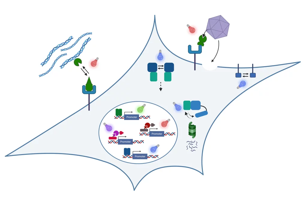
Biosensors
We integrate natural and engineered molecular sensors for drugs, metabolites or nucleic acids into suitable readout formats for the fast and sensitive quantification of such substances. Together with collaboration partners, we develop biosensor systems for different application fields:
Open Positions
We are always excited to meet curious and creative scientists passionate about synthetic biology, optogenetics, and engineered living materials. If you would like to shape the future of biobased and living materials with us, we warmly welcome your spontaneous application for a PhD thesis or Postdoc position!
Projects and Partners
We perform collaborative research in materials-oriented synthetic biology within interdisciplinary research consortia
STEADY
Within the ERC Advanced Grant STEADY, we develop concepts for dynamically controlling the properties of engineered living materials by advanced synthetic genetic circuits.
LoopOfFun
We coordinate the European Innovation Council (EIC)-funded consortium LoopOfFun in which we aim at developing a platform for the rapid development of industry-scale, one-step, simple casting-based manufacturing processes for fungal mycelia composites. We jointly work towards this goal with our consortium partners:
- Prof. Roman Jerala, National Institute of Chemistry, Ljubljana, Slovenia
- Dr. Achim Weber, Fraunhofer IGB, Stuttgart, Germany
- Prof. Arnold Driessen, University of Groningen, The Netherlands
- Carlotta Borgato and Jan Boelen, Atelier LUMA, Arles, France
DELIVER
In the project DELIVER funded by the Carl-Zeiss-Foundation, we collaborate towards the data-driven engineering of sustainable living materials. We combine synthetic biology with materials sciences and data-driven approaches to design bio-based composite materials with custom-tailored structural properties for construction applications. Within deliver, we collaborate with the following partners:
- Prof. Thomas Speck, University of Freiburg, Germany
- Dr. Clemens Kreutz, University Hospital Freiburg, Germany
BILLARD
We coordinate the BILLARD project funded by the Federal Ministry of Education and Research (BMBF) within the funding line “Biologization of Technology”, we collaborate with PD Dr. Felicitas Bucher from the Clinic of Ophtamology at the University Hospital Freiburg on the development of novel intraocular drug delivery devices.
CIBSS – Centre for Integrative Biological Signalling Studies
We are member of the Cluster of Excellence CIBSS in which we perform research on novel optogenetic technologies to control signaling reactions in mammalian cells. We mainly collaborate with Prof. Dr. Jens Timmer on the model-based design of synthetic biological switches and networks and with Prof. Dr. Wolfgang Schamel on controlling immunological processes such as T cell activation via optogenetics.
Publications
Smith, R. W. | Helwig, B. | Westphal, A. H. | Pel, E. | Hörner, M. | Beyer, H. M. | Samodelov, S. L. | Weber, Wilfried | Zurbriggen, M. D. | Borst, J. W. | Fleck, C.
DOI:
Background: Obtaining accurate estimates of biological or enzymatic reaction rates is critical in understanding the design principles of a network and how biological processes can be experimentally manipulated on demand. In many cases experimental limitations mean that some enzymatic rates cannot be measured directly, requiring mathematical algorithms to estimate them. Here, we describe a methodology that calculates rates at which light-regulated proteins switch between conformational states. We focus our analysis on the phytochrome family of photoreceptors found in cyanobacteria, plants and many optogenetic tools. Phytochrome proteins change between active (P A) and inactive (P I) states at rates that are proportional to photoconversion cross-sections and influenced by light quality, light intensity, thermal reactions and dimerisation. This work presents a method that can accurately calculate these photoconversion cross-sections in the presence of multiple non-light regulated reactions. Results: Our approach to calculating the photoconversion cross-sections comprises three steps: i) calculate the thermal reversion reaction rate(s); ii) develop search spaces from which all possible sets of photoconversion cross-sections exist, and iii) estimate extinction coefficients that describe our absorption spectra. We confirm that the presented approach yields accurate results through the use of simulated test cases. Our test cases were further expanded to more realistic scenarios where noise, multiple thermal reactions and dimerisation are considered. Finally, we present the photoconversion cross-sections of an Arabidopsis phyB N-terminal fragment commonly used in optogenetic tools. Conclusions: The calculation of photoconversion cross-sections has implications for both photoreceptor and synthetic biologists. Our method allows, for the first time, direct comparisons of photoconversion cross-sections and response speeds of photoreceptors in different cellular environments and synthetic tools. Due to the generality of our procedure, as shown by the application to multiple test cases, the photoconversion cross-sections and quantum yields of any photoreceptor might now, in principle, be obtained. © 2016 The Author(s).
Wagner, H. J. | Sprenger, A. | Rebmann, B. | Weber, Wilfried
DOI:
One key aspect of synthetic biology is the development and characterization of modular biological building blocks that can be assembled to construct integrated cell-based circuits performing computational functions. Likewise, the idea of extracting biological modules from the cellular context has led to the development of in vitro operating systems. This principle has attracted substantial interest to extend the repertoire of functional materials by connecting them with modules derived from synthetic biology. In this respect, synthetic biological switches and sensors, as well as biological targeting or structure modules, have been employed to upgrade functions of polymers and solid inorganic material. The resulting systems hold great promise for a variety of applications in diagnosis, tissue engineering, and drug delivery. This review reflects on the most recent developments and critically discusses challenges concerning in vivo functionality and tolerance that must be addressed to allow the future translation of such synthetic biology-upgraded materials from the bench to the bedside. © 2016 Elsevier B.V.
Beyer, H. M. | Gonschorek, P. | Samodelov, S. L. | Meier, M. | Weber, Wilfried | Zurbriggen, M. D.
DOI:
Assembly cloning is increasingly replacing conventional restriction enzyme and DNAligase-dependent cloning methods for reasons of efficiency and performance. Here, we describe AQUA (advanced quick assembly), a simple and versatile seamless assembly cloning approach. We demonstrate the applicability and versatility of AQUA Cloning in selected proof-of-principle applications including targeted insertion-, deletion- and sitedirected point-mutagenesis, and combinatorial cloning. Furthermore, we show the one pot de novo assembly of multiple DNA fragments into a single circular plasmid encoding a complex light- and chemically-regulated Boolean A NIMPLY B logic operation. AQUA Cloning harnesses intrinsic in vivo processing of linear DNA fragments with short regions of homology of 16 to 32 bp mediated by Escherichia coli. It does not require any kits, enzymes or preparations of reagents and is the simplest assembly cloning protocol to date. © 2015 Beyer et al. This is an open access article distributed under the terms of the Creative Commons Attribution License, which permits unrestricted use, distribution, and reproduction in an medium, provided the original author and source are credited.
Beyer, H. M. | Juillot, S. | Herbst, K. | Samodelov, S. L. | Müller, K. | Schamel, W. W. | Römer, W. | Schäfer, E. | Nagy, F. | Strähle, U. | Weber, Wilfried | Zurbriggen, M. D.
DOI:
Protein trafficking in and out of the nucleus represents a key step in controlling cell fate and function. Here we report the development of a red light-inducible and far-red light-reversible synthetic system for controlling nuclear localization of proteins in mammalian cells and zebrafish. First, we synthetically reconstructed and validated the red light-dependent Arabidopsis phytochrome B nuclear import mediated by phytochrome-interacting factor 3 in a nonplant environment and support current hypotheses on the import mechanism in planta. On the basis of this principle we next regulated nuclear import and activity of target proteins by the spatiotemporal projection of light patterns. A synthetic transcription factor was translocated into the nucleus of mammalian cells and zebrafish to drive transgene expression. These data demonstrate the first in vivo application of a plant phytochrome-based optogenetic tool in vertebrates and expand the repertoire of available light-regulated molecular devices. © 2015 American Chemical Society.
Beyer, H. M. | Naumann, S. | Weber, Wilfried | Radziwill, G.
DOI:
Molecular signals are sensed by their respective receptors and information is transmitted and processed by a sophisticated intracellular network controlling various biological functions. Optogenetic tools allow the targeting of specific signaling nodes for a precise spatiotemporal control of downstream effects. These tools are based on photoreceptors such as phytochrome B (PhyB), cryptochrome 2, or light-oxygen-voltage-sensing domains that reversibly bind to specific interaction partners in a light-dependent manner. Fusions of a protein of interest to the photoreceptor or their interaction partners may enable the control of the protein function by light-mediated dimerization, a change of subcellular localization, or due to photocaging/-uncaging of effectors. In this review, we summarize the photoreceptors and the light-based mechanisms utilized for the modulation of signaling events in mammalian cells focusing on non-neuronal applications. We discuss in detail optogenetic tools and approaches applied to control signaling events mediated by second messengers, Rho GTPases and growth factor-triggered signaling cascades namely the RAS/RAF and phosphatidylinositol-3-kinase pathways. Applying the latest generation of optogenetic tools allows to control cell fate decisions such as proliferation and differentiation or to deliver therapeutic substances in a spatiotemporally controlled manner. Optogenetic tools enable the spatiotemporal control of the function of genetically encoded proteins by light. The authors discuss optogenetic tools and approaches applied to control signaling events mediated by second messengers, Rho GTPases and signaling cascades namely the RAS/RAF and PI3K/AKT pathways. Applying the latest generation of optogenetic tools allows to control cell fate decisions such as proliferation and differentiation or to deliver therapeutic substances in a spatiotemporally-controlled manner. © 2014 WILEY-VCH Verlag GmbH & Co. KGaA, Weinheim.
Ketterer, S. | Fuchs, D. | Weber, Wilfried | Meier, M.
DOI:
Fluorogenic RNAs that are based on the complex formed by 3,5-difluoro-4-hydroxybenzylidene imidazolinone (DFHBI) derivatives and the RNA aptamer named Spinach were used to engineer a new generation of in vitro and in vivo sensors for bioanalytics. With the resolved crystal structure of the RNA/small molecule complex, the engineering map becomes available, but comprehensive information regarding the thermodynamic profile of the molecule is missing. Here, we reconstructed the full thermodynamic binding and stability landscapes between DFHBI and a truncated sequence of first-generation Spinach. For this purpose, we established a systematic screening procedure for single- and double-point mutations on a microfluidic large-scale integrated chip platform for 87-nt long RNAs. The thermodynamic profile with single base resolution was used to engineer an improved fluorogenic spinach generation via a directed rather than evolutional approach. © The Author(s) 2015. Published by Oxford University Press on behalf of Nucleic Acids Research.
Ketterer, S. | Hövermann, D. | Guebeli, R. J. | Bartels-Burgahn, F. | Riewe, D. | Altmann, T. | Zurbriggen, M. D. | Junker, B. | Weber, Wilfried | Meier, M.
DOI:
DOI:Kolar, K. | Wischhusen, H. M. | Müller, K. | Karlsson, M. | Weber, Wilfried | Zurbriggen, M. D.
DOI:
Background: Multicellular organisms depend on the exchange of information between specialized cells. This communication is often difficult to decipher in its native context, but synthetic biology provides tools to engineer well-defined systems that allow the convenient study and manipulation of intercellular communication networks. Results: Here, we present the first mammalian synthetic network for reciprocal cell-cell communication to compute the border between a sender/receiver and a processing cell population. The two populations communicate via Ltryptophan and interleukin-4 to highlight the population border by the production of a fluorescent protein. The sharpness of that visualized edge can be adjusted by modulating key parameters of the network. Conclusions: We anticipate that this network will on the one hand be a useful tool to gain deeper insights into the mechanisms of tissue formation in nature and will on the other hand contribute to our ability to engineer artificial tissues. © 2015 Kolar et al.
Lienemann, P. S. | Devaud, Y. R. | Reuten, R. | Simona, B. R. | Karlsson, M. | Weber, Wilfried | Koch, M. | Lutolf, M. P. | Milleret, V. | Ehrbar, M.
DOI:
The perivascular niche is a complex microenvironment containing mesenchymal stem cells (MSCs), among other perivascular cells, as well as temporally organized biochemical and biophysical gradients. Due to a lack of conclusive phenotypic markers, MSCs' identity, heterogeneity and function within their native niche remain poorly understood. The in vitro reconstruction of an artificial three-dimensional (3D) perivascular niche would offer a powerful alternative to study MSC behavior under more defined conditions. To this end, we here present a poly(ethylene glycol)-based in vitro model that begins to mimic the spatiotemporally controlled presentation of biological cues within the in vivo perivascular niche, namely a stably localized platelet-derived growth factor B (PDGF-BB) gradient. We show that 3D-encapsulated MSCs respond to soluble PDGF-BB by proliferation, spreading, and migration in a dose-dependent manner. In contrast, the exposure of MSCs to 3D matrix-tethered PDGF-BB gradients resulted in locally restricted morphogenetic responses, much as would be expected in a native perivascular niche. Thus, the herein presented artificial perivascular niche model provides an important first step towards modeling the role of MSCs during tissue homeostasis and regeneration. © 2015 The Royal Society of Chemistry.
Metzger, S. | Lienemann, P. S. | Ghayor, C. | Weber, Wilfried | Martin, I. | Weber, F. E. | Ehrbar, M.
DOI:
The in vitro formation of physiologically relevant engineered tissues is still limited by the availability of adequate growth-factor-presenting cell-instructive biomaterials, allowing simultaneous and three-dimensionally localized differentiation of multiple tissue progenitor cells. Together with ever improving technologies such as microfluidics, printing, or lithography, these biomaterials could provide the basis for generating provisional cellular constructs, which can differentiate to form tissue mimetics. Although state-of-the-art biomaterials are endowed with sophisticated modules for time- and space-controlled positioning and release of bioactive molecules, reports on 3D arrangements of differentiation-inducing growth factors are scarce. This paper describes the stable and localized immobilization of biotinylated bioactive molecules to a modular, Factor XIII-cross-linked poly(ethylene glycol) hydrogel platform using a genetically engineered streptavidin linker. Linker incorporation is demonstrated by Western blot, and streptavidin functionality is confirmed by capturing biotinylated alkaline phosphatase (ALP). After optimizing bone morphogenetic protein 2 (BMP-2) biotinylation, streptavidin-modified hydrogels are able to bind and present bioactive BMP-2-biotin. Finally, with this immobilization scheme for BMP-2, the specific osteogenic differentiation of mesenchymal stem cells is demonstrated by inducing ALP expression in confined 3D areas. In future, this platform together with other affinity-based strategies will be useful for the local incorporation of various growth factors for engineering cell-responsive constructs. © 2014 WILEY-VCH Verlag GmbH & Co. KGaA, Weinheim.