

We engineer cells and materials that communicate and process information through synthetic biology
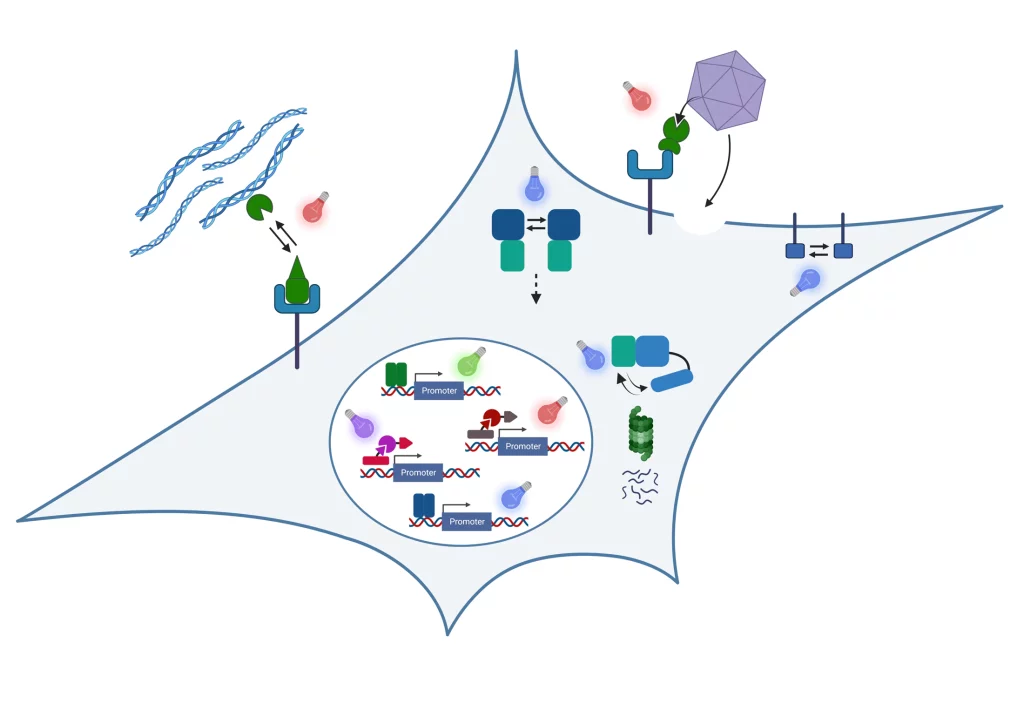
Our inspiration is the ability of organisms and the materials they are made of to adapt to dynamic environmental conditions. Plants adapt growth to light conditions; bacteria develop resistance against antibiotics or bones get stronger when exercised. The basis for this ability to adapt is a fascinating information processing machinery of the organisms: Environmental conditions are captured by molecular sensors, then the signals are processed and integrated with genetic programs to finally yield a targeted response.
In our research, we engineer nature’s molecular sensing, processing, and actuation machinery in order to precisely control the function and properties of cells and materials. We apply these newly developed technologies in different fields of fundamental and applied research.

Team Members


Research
Stimulus-responsive and Information-processing (living) Materials

We develop and apply stimulus-responsive and information-processing biohybrid polymer materials. To this aim, we functionally couple synthetic biological molecular sensors and switches to polymer materials. By wiring these switches according to topologies inspired by electronic circuits, we engineer materials that perform fundamental computational operations. Examples of our work include:
- We engineered a hydrogel based on a bacteria-derived photoreceptor which allows the light-responsive, fully reversibly tuning of its mechanical properties. We applied this hydrogel as extracellular matrix to analyze the impact of dynamic mechanical environments on transcriptome-wide responses in mesenchymal stem cells or on the migration of T-lymphocytes.
See Hörner et al. Advanced Materials 2019 - We integrated synthetic biological switches with polymer materials into a circuit inspired by an electronic counter. The resulting material system was able to count the number of input light pulses and to release different output as a function of the number of light pulses detected. We applied this system to sequentially release different biocatalysts to drive a two-step biochemical reaction.
See Beyer et al., Advanced Materials 2018 - We developed PenTag, a protein tag for the spontaneous, covalent coupling of proteins to ampicillin-functionalized molecules such as dyes, polymers, or solid supports. Based on this strategy, we engineered and assembled material modules to function as encoder for processing different combinations of biochemical input stimuli.
See Mohsenin et al., Advanced Functional Materials 2024 - By engineering modular protease-based switches that can either be activated or repressed, we develop information-processing biohybrid circuits that process binary biomolecular information according to a circuit inspired by electronic decoders. Such circuits can be applied to process and interpret biochemical sensor information for advanced diagnostic applications.
See Mohsenin et al., Advanced Materials 2024
Molecular optogenetics to control cell fate and function
We develop and apply molecular optogenetic tools to control cell fate and function with unprecedented spatial and temporal precision in a dose-dependent and highly specific manner. To this aim, we engineer plant- and bacteria-derived photoreceptors and functionally couple them to proteins involved in cell signaling and gene expression. Examples of our work include:
- Light-inducible formation of liquid or gel-like transcription factor condensates in mammalian cells and mice. We demonstrate that liquid “transcription factor droplets” show a several-fold higher activity in inducing transgene expression compared to native transcription factors. Further, gel-like transcription factor condensates were shown to correlate with decreased transcriptional activation thus providing a materials-based layer of controlling gene expression.
See Schneider et al., Science Advances 2021 and Fischer et al., Small 2024 - Light-guided adeno-associated viral (AAV) vectors. We engineered a light-responsive tropism into AAVs which allows us to selectively transfer genetic information into single cells or to transduce different cells within one culture with different transgenes.
See Hörner et al., Science Advances 2021
Our group is running www.optobase.org, the most comprehensive database on molecular optogenetics. Have a look and discover the amazing opportunities in controlling biology with light!
Biosensors
We integrate natural and engineered molecular sensors for drugs, metabolites or nucleic acids into suitable readout formats for the fast and sensitive quantification of such substances. Together with collaboration partners, we develop biosensor systems for different application fields:
Open Positions
We are always excited to meet curious and creative scientists passionate about synthetic biology, optogenetics, and engineered living materials. If you would like to shape the future of biobased and living materials with us, we warmly welcome your spontaneous application for a PhD thesis or Postdoc position!
Projects and Partners
We perform collaborative research in materials-oriented synthetic biology within interdisciplinary research consortia
STEADY
Within the ERC Advanced Grant STEADY, we develop concepts for dynamically controlling the properties of engineered living materials by advanced synthetic genetic circuits.
LoopOfFun
We coordinate the European Innovation Council (EIC)-funded consortium LoopOfFun in which we aim at developing a platform for the rapid development of industry-scale, one-step, simple casting-based manufacturing processes for fungal mycelia composites. We jointly work towards this goal with our consortium partners:
- Prof. Roman Jerala, National Institute of Chemistry, Ljubljana, Slovenia
- Dr. Achim Weber, Fraunhofer IGB, Stuttgart, Germany
- Prof. Arnold Driessen, University of Groningen, The Netherlands
- Carlotta Borgato and Jan Boelen, Atelier LUMA, Arles, France
DELIVER
In the project DELIVER funded by the Carl-Zeiss-Foundation, we collaborate towards the data-driven engineering of sustainable living materials. We combine synthetic biology with materials sciences and data-driven approaches to design bio-based composite materials with custom-tailored structural properties for construction applications. Within deliver, we collaborate with the following partners:
- Prof. Thomas Speck, University of Freiburg, Germany
- Dr. Clemens Kreutz, University Hospital Freiburg, Germany
BILLARD
We coordinate the BILLARD project funded by the Federal Ministry of Education and Research (BMBF) within the funding line “Biologization of Technology”, we collaborate with PD Dr. Felicitas Bucher from the Clinic of Ophtamology at the University Hospital Freiburg on the development of novel intraocular drug delivery devices.
CIBSS – Centre for Integrative Biological Signalling Studies
We are member of the Cluster of Excellence CIBSS in which we perform research on novel optogenetic technologies to control signaling reactions in mammalian cells. We mainly collaborate with Prof. Dr. Jens Timmer on the model-based design of synthetic biological switches and networks and with Prof. Dr. Wolfgang Schamel on controlling immunological processes such as T cell activation via optogenetics.
Publications
Mühlhäuser, W. W. D. | Fischer, A. | Weber, Wilfried | Radziwill, G.
DOI:
Cells receive many different environmental clues to which they must adapt accordingly. Therefore, a complex signal transduction network has evolved. Cellular signal transduction is a highly dynamic process, in which the specific outcome is a result of the exact spatial and temporal resolution of single sub-events. While conventional techniques, like chemical inducer systems, have led to a sound understanding of the architecture of signal transduction pathways, the spatiotemporal aspects were often impossible to resolve. Optogenetics, based on genetically encoded light-responsive proteins, has the potential to revolutionize manipulation of signal transduction processes. Light can be easily applied with highest precision and minimal invasiveness. This review focuses on examples of optogenetic systems which were generated and applied to manipulate non-neuronal mammalian signaling processes at various stages of signal transduction, from cell membrane through cytoplasm to nucleus. Further, the future of optogenetic signaling will be discussed. © 2016 Elsevier B.V.
Mühlhäuser, W. W. D. | Hörner, M. | Weber, Wilfried | Radziwill, G.
DOI:
Optogenetic approaches enable the control of biological processes in a time- and space-resolved manner. These light-based methods are noninvasive and by using light as sole activator minimize side effects in contrast to chemical inducers. Here, we provide a protocol for the targeted control of the activity of protein kinases in mammalian cells based on the photoreceptor cryptochrome 2 (CRY2) of Arabidopsis thaliana and its interaction partner CIB1. Blue light (450 nm)-induced binding of CRY2 to CIB1 allows the recruitment of a chimeric cytosolic protein kinase AKT1 to the plasma membrane accompanied with stimulation of its kinase activity. This protocol comprises the transient and stable implementation of the light-regulated system into mammalian cells and its stimulation by blue light-emitting diodes (450 nm) irradiation as well as analysis of the light-activated AKT1. © Springer Science+Business Media LLC 2017.
Chatelle, C. V. | Hövermann, D. | Müller, A. | Wagner, H. J. | Weber, Wilfried | Radziwill, G.
DOI:
Here, we applied optoRAF, an optogenetic tool for light-controlled clustering and activation of RAF proteins that mimics the natural occurring RAS-mediated dimerization. This versatile tool allows studying the effect on BRAF and CRAF homodimer- as well as heterodimer-induced RAF signaling. Vemurafenib and dabrafenib are two clinically approved inhibitors for BRAF that efficiently suppress the kinase activity of oncogenic BRAF (V600E). However in wild-type BRAF expressing cells, BRAF inhibitors can exert paradoxical activation of wild-type CRAF. Using optoRAF, vemurafenib was identified as paradoxical activator of BRAF and CRAF homo- and heterodimers. Dabrafenib enhanced activity of light-stimulated CRAF at low dose and inhibited CRAF signaling at high dose. Moreover, dabrafenib increased the protein level of CRAF proteins but not of BRAF proteins. Increased CRAF levels correlate with elevated RAF signaling in a dabrafenib-dependent manner, independent of light activation.
Dincer, C. | Kling, A. | Chatelle, C. | Armbrecht, L. | Kieninger, J. | Weber, Wilfried | Urban, G. A.
DOI:
Here, we present a novel approach to increase the degree of miniaturization as well as the sensitivity of biosensor platforms by the optimization of microfluidic stop-flow techniques independent of the applied detection technique (e.g. electrochemical or optical). The readout of the labeled bioassays, immobilized in a microfluidic channel, under stop-flow conditions leads to a rectangular shaped peak signal. Data evaluation using the peak height allows for a high level miniaturization of the channel geometries. To study the main advantages and limitations of this method by numerical simulations, a universally applicable model system is introduced for the first time. Consequently, proof-of-principle experiments were successfully performed with standard and miniaturized versions of an electrochemical biosensor platform utilizing a repressor protein-based assay for tetracycline antibiotics. Herein, the measured current peak heights are the same despite the sextuple reduction of the channel dimensions. Thus, this results in a 22-fold signal amplification compared to the constant flow measurements in the case of the miniaturized version. © 2016 The Royal Society of Chemistry.
Fischer, A. | Warscheid, B. | Weber, Wilfried | Radziwill, G.
DOI:
Scaffold proteins such as the multidomain protein CNK1 orchestrate the signalling network by integrating and controlling the underlying pathways. Using an optogenetic approach to stimulate CNK1 uncoupled from upstream effectors, we identified selective clusters of CNK1 that either stimulate RAF-MEK-ERK or AKT signalling depending on the light intensity applied. OptoCNK1 implemented in MCF7 cells induces differentiation at low light intensity stimulating ERK activity whereas stimulation of AKT signalling by higher light intensity promotes cell proliferation. CNK1 clustering in response to increasing EGF concentrations revealed that CNK1 binds to RAF correlating with ERK activation at low EGF dose. At higher EGF dose active AKT binds to CNK1 and phosphorylates and inhibits RAF. Knockdown of CNK1 protects CNK1 from this AKT/RAF crosstalk. In C2 skeletal muscle cells CNK1 expression is induced with the onset of differentiation. Hence, AKT-bound CNK1 counteracts ERK stimulation in differentiated but not in proliferating cells. Ectopically expressed CNK1 facilitates C2 cell differentiation and knockdown of CNK1 impaired the transcriptional network underlying C2 cell differentiation. Thus, CNK1 expression, CNK1 clustering and the thereto related differential signalling processes decide on proliferation and differentiation in a cell type-and cell stage-dependent manner by orchestrating AKT and RAF signalling. © 2016 The Author(s).
Juillot, S. | Beyer, H. M. | Madl, J. | Weber, Wilfried | Zurbriggen, M. D. | Römer, W.
DOI:
One major regulatory mechanism in cell signalling is the spatiooral control of the localization of signalling molecules. We synthetically designed an entire cell signalling pathway, which allows controlling the transport of signalling molecules from the plasma membrane to the nucleus, by using light and small molecules. © The Royal Society of Chemistry 2016.
Kling, A. | Chatelle, C. | Armbrecht, L. | Qelibari, E. | Kieninger, J. | Dincer, C. | Weber, Wilfried | Urban, G.
DOI:
The excessive use of antibiotics in human and veterinary medicine causes the emergence of multidrug resistant bacteria. In this context, the surveillance of many different antibiotics provokes a worldwide challenge. Hence, fast and versatile multianalyte single-use biosensors are of increasing interest for many fields such as medical analysis or environmental and food control. Here we present a microfluidic platform enabling the electrochemical readout of up to eight enzyme-linked assays (ELAs), simultaneously. To demonstrate the applicability of this platform for the surveillance and monitoring of antibiotics, we used highly sensitive biomolecular sensor systems for the simultaneous detection of two commonly employed antibiotic classes tetracycline and streptogramin. Thus, microfluidic channel networks are designed, comprising distinct numbers of immobilization sections with a very low volume of 680 nL each. These passively metered sections can be actuated separately for an individual assay procedure. The limits of detection (LOD) are determined, with high precision, to 6.33 and 9.22 ng mL-1 for tetracycline and pristinamycin, respectively. The employed channel material, dry film photoresist (DFR), allows an easy storage of preimmobilized assays with a shelf life of at least 3 months. Multianalyte measurements in a complex medium are demonstrated by the simultaneous detection of both antibiotics in spiked human plasma within a sample-to-result time of less than 15 min. © 2016 American Chemical Society.
Metzger, S. | Blache, U. | Lienemann, P. S. | Karlsson, M. | Weber, F. E. | Weber, Wilfried | Ehrbar, M.
DOI:
Engineering in vitro tissue mimetics that resemble the corresponding living tissues requires the 3D arrangement of tissue progenitor cells and their differentiation by localized growth factor (GF) signaling cues. Recent technological advances open a large field of possibilities for the creation of complex GF arrangements. Additionally, cell-instructive biomaterials, which bind GFs by various mechanisms and release them with different kinetics depending on binding affinity, have become available. This paper describes the development of a matrix metalloproteinase (MMP)-degradable streptavidin-based linker module, which allows the release of immobilized GFs from synthetic biomimetic poly(ethylene glycol) hydrogels independently of the hydrogel degradation. The MMP-sensitive streptavidin linker is shown to efficiently bind biotinylated molecules, and as proof of concept, bone morphogenetic protein-2 (BMP-2) delivery via the MMP-degradable linker is used to induce osteogenic differentiation in C2C12 cells and mesenchymal stem cells. The results show a significantly increased net effect of proteolytically releasable BMP-2 in comparison to stably immobilized and soluble BMP-2. This study indicates that a GF delivery system directly responsive to cellular activity can have important implications for the synthesis of tissue mimetics and regenerative medicine, as it can influence the availability, the localization of effects, as well as efficacy of employed GFs. (Figure presented.). © 2016 WILEY-VCH Verlag GmbH & Co. KGaA, Weinheim
Ochoa-Fernandez, R. | Samodelov, S. L. | Brandl, S. M. | Wehinger, E. | Müller, K. | Weber, Wilfried | Zurbriggen, M. D.
DOI:
Optogenetic tools to control gene expression have many advantages over the classical chemically inducible systems, overcoming intrinsic limitations of chemical inducers such as solubility, diffusion, and cell toxicity. They offer an unmatched spatiotemporal resolution and permit quantitative and noninvasive control of the gene expression. Here we describe a protocol of a synthetic light-inducible system for the targeted control of gene expression in plants based on the plant photoreceptor phytochrome B and one of its interacting factors (PIF6). The synthetic toggle switch system is in the ON state when plant protoplasts are illuminated with red light (660 nm) and can be returned to the OFF state by subsequent illumination with farred light (760 nm). In this protocol, the implementation of a red light-inducible expression system in plants using Light-Emitting Diode (LED) illumination boxes is described, including the isolation and transient transformation of plant protoplasts from Arabidopsis thaliana and Nicotiana tabacum . © Springer Science+Business Media New York 2016.
Samodelov, S. L. | Beyer, H. M. | Guo, X. | Augustin, M. | Jia, K. P. | Baz, L. | Ebenhöh, O. | Beyer, P. | Weber, Wilfried | Al-Babili, S. | Zurbriggen, M. D.
DOI:
Strigolactones are key regulators of plant development and interaction with symbiotic fungi; however, quantitative tools for strigolactone signaling analysis are lacking. We introduce a genetically encoded hormone biosensor used to analyze strigolactone-mediated processes, including the study of the components involved in the hormone perception/signaling complex and the structural specificity and sensitivity of natural and synthetic strigolactones in Arabidopsis, providing quantitative insights into the stereoselectivity of strigolactone perception. Given the high specificity, sensitivity, dynamic range of activity, modular construction, ease of implementation, and wide applicability, the biosensor StrigoQuant will be useful in unraveling multiple levels of strigolactone metabolic and signaling networks. © 2016 The Author.